Seborrhoeic
Keratosis.
Just a gentle reminder that our skin is getting a little older.

Just a gentle reminder that our skin is getting a little older.
The full word is a mouthful for such a common harmless growth, so let’s shorten it to ‘SK.’
SK is a very common warty spot that occurs as we get older. SK is mainly made up of keratinocytes, the predominant cells of the upper layer of the skin (epidermis). Keratinocytes produce keratin which is found in scale.
Hearing the name SK is an expected part of a routine skin check.
SK’s should be famous – Most people over 60 years of age have at least 1 seborrhoeic keratosis.
They are present in both males and females and typically start appearing after the age of 40.
There is a genetic predisposition to have multiple lesions.
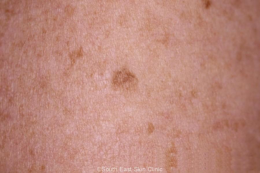
The appearance of seborrhoeic keratoses is extremely diverse.
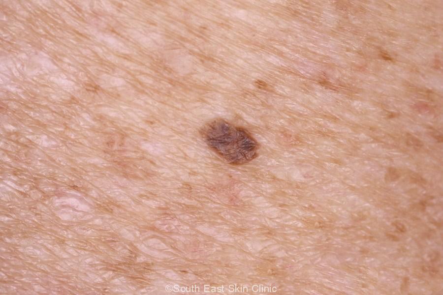
An SK is generally scaly and brown in colour (light to dark). The colour can vary from pink through to black, and they generally range in size from 1-3cm in diameter.
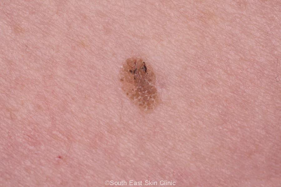
Initially, the lesion is flat, gradually becoming raised from the skin surface – by which time it appears ‘stuck on.’ The surface is dull, warty, or waxy.
The pink or light brown SKs are more likely to be dull, whilst the darker brown ones are typically waxy. You can see this in the gallery images.
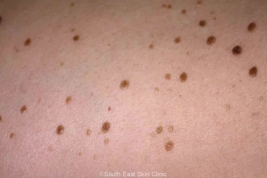
The face and trunk are commonly affected areas however they can appear anywhere on the body. It is not uncommon to have multiple SKs.
Dermatoscopy of SK will show characteristic features, avoiding the need for a biopsy – but not always.
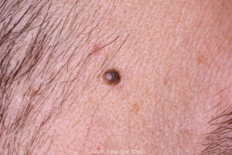
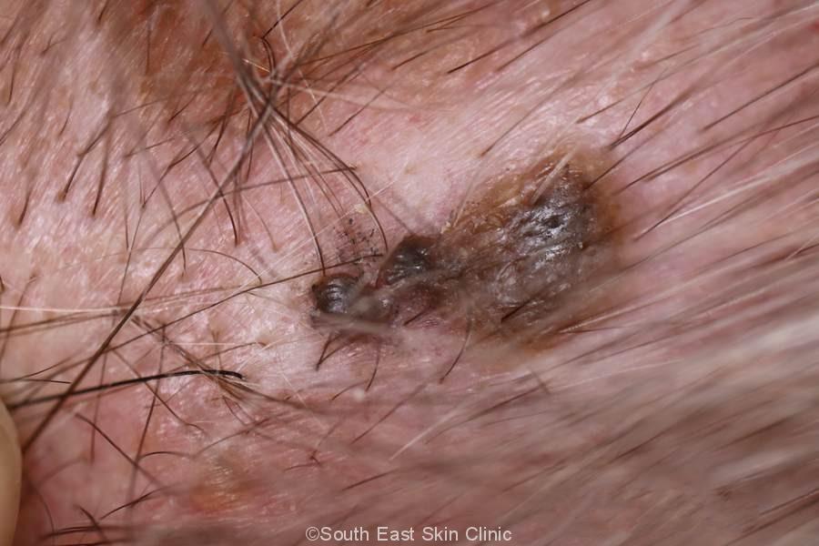
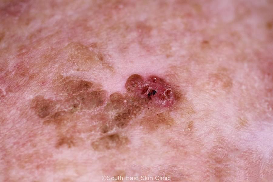
A pitfall is ‘melanoacanthomatous melanoma’ – in other words, a melanoma that looks like an SK.
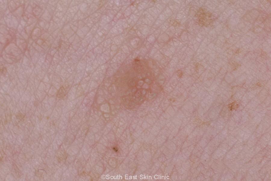
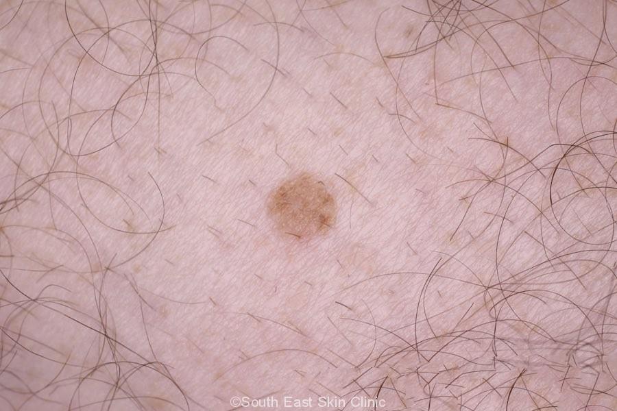
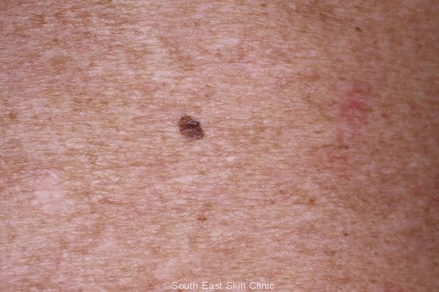
Flat Seborrhoeic keratosis
Please click on the images for details.
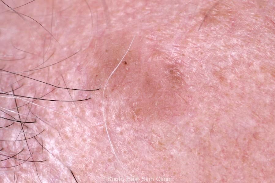
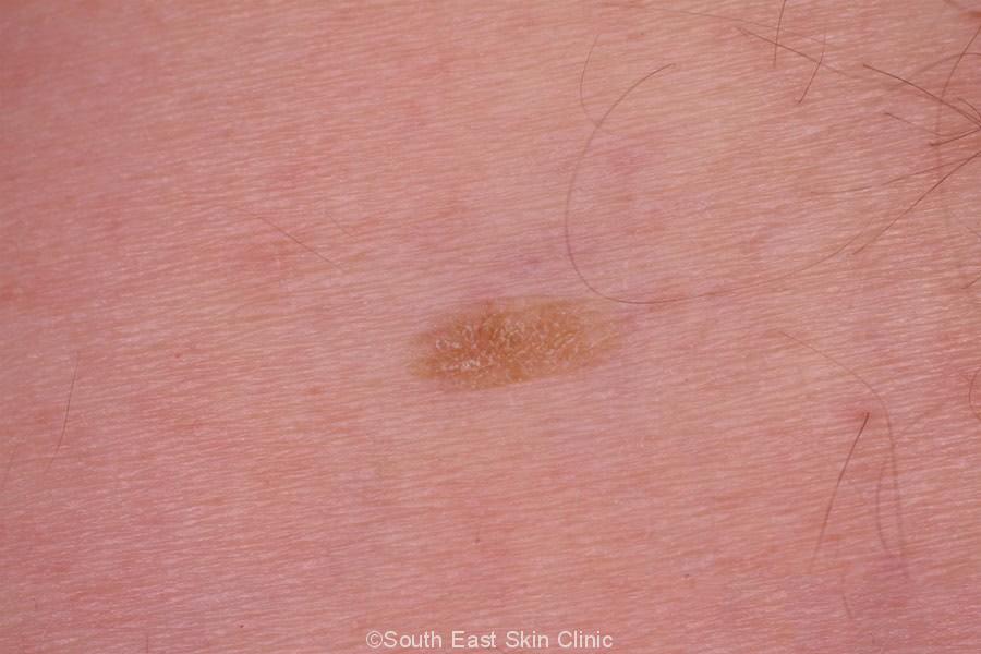
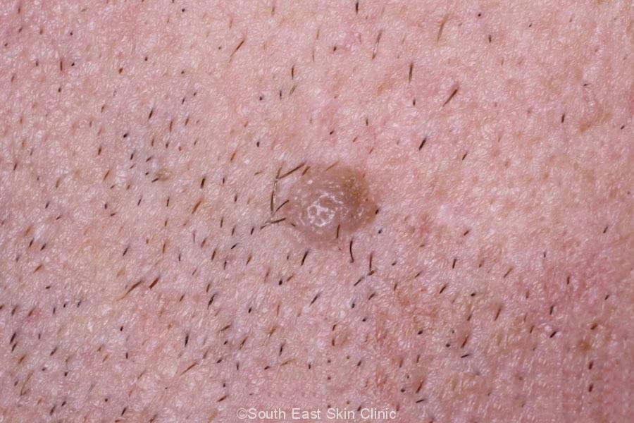
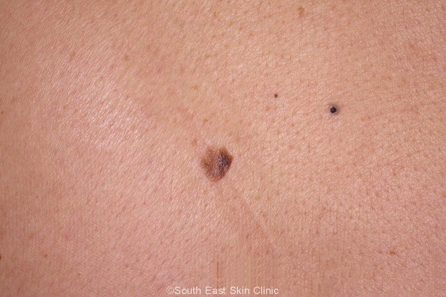
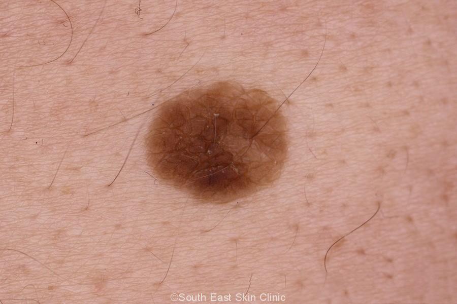
Pink Seborrhoeic keratosis
Please click on the images for details.
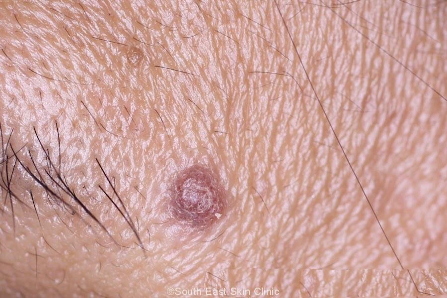
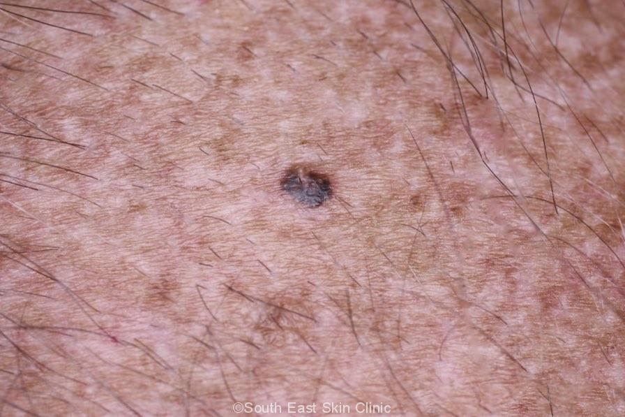
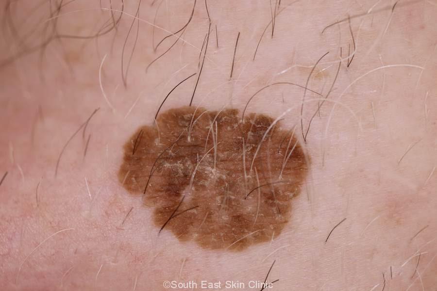
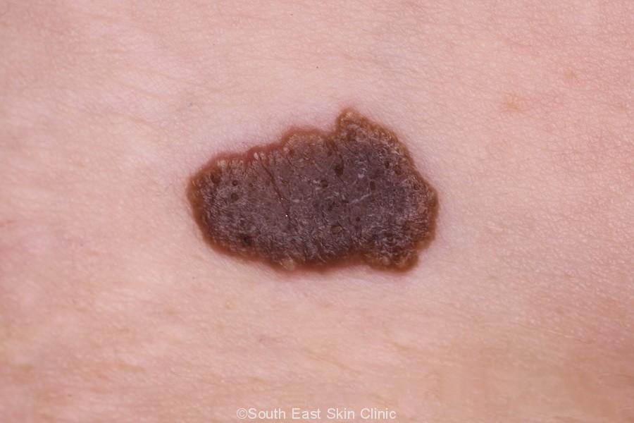
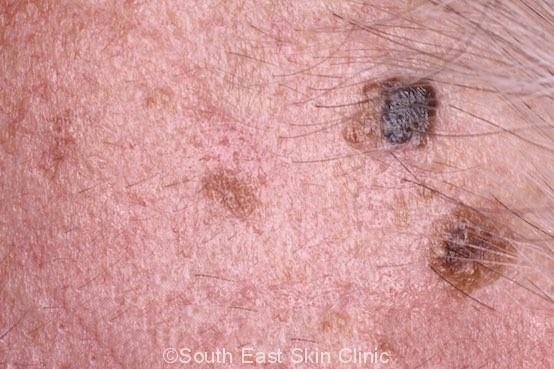
Dark Seborrhoeic keratosis may be confused with melanoma
Please click on the images for details.
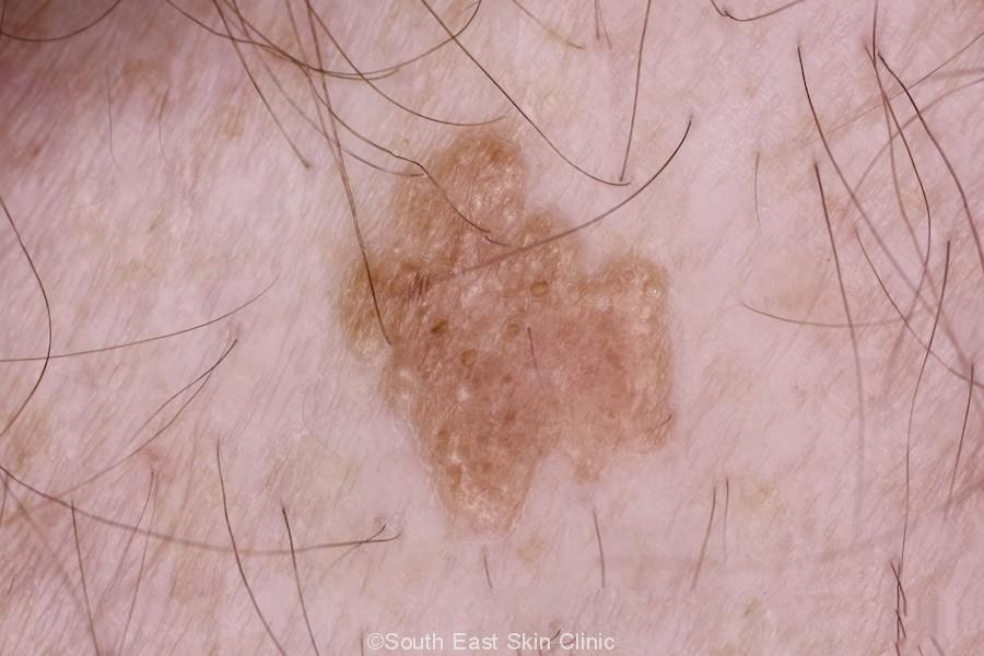
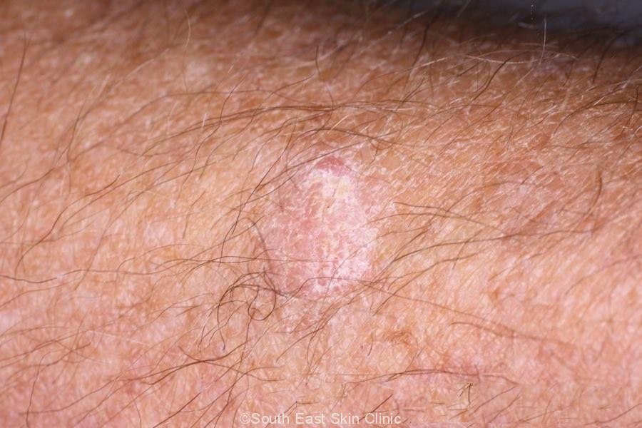
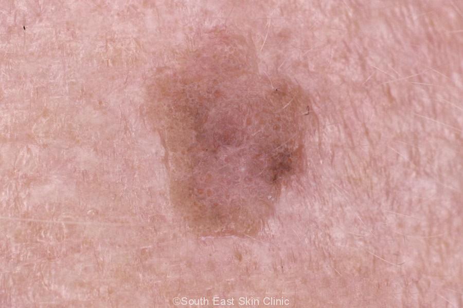
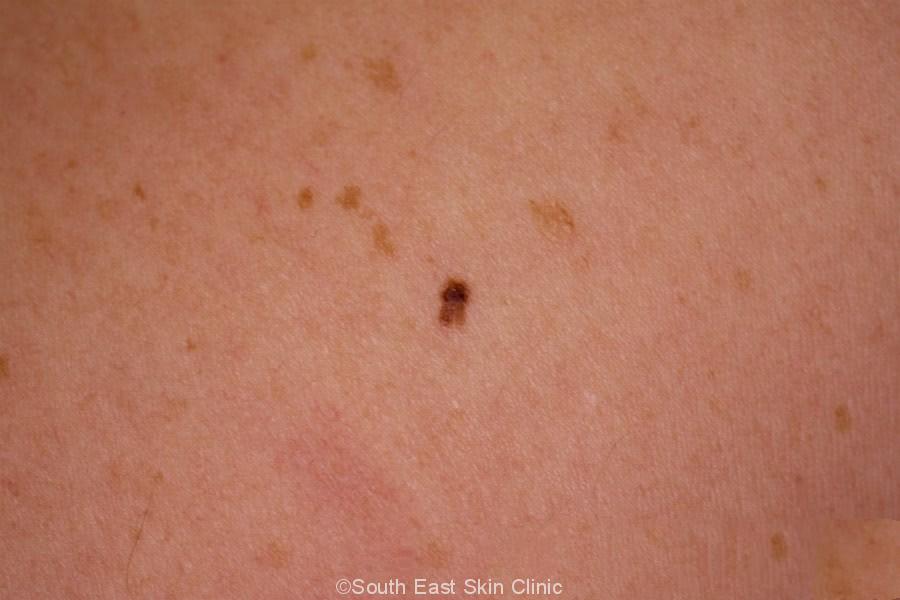
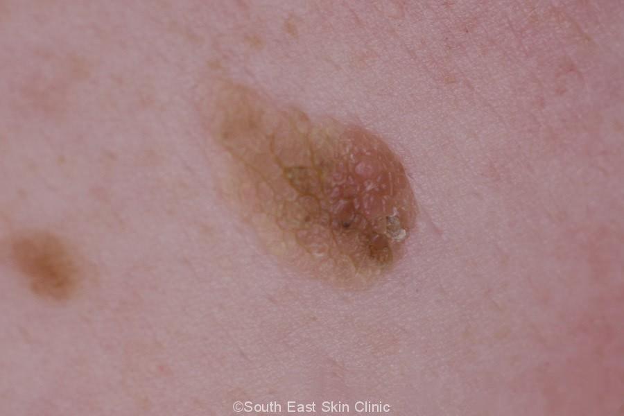
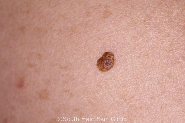
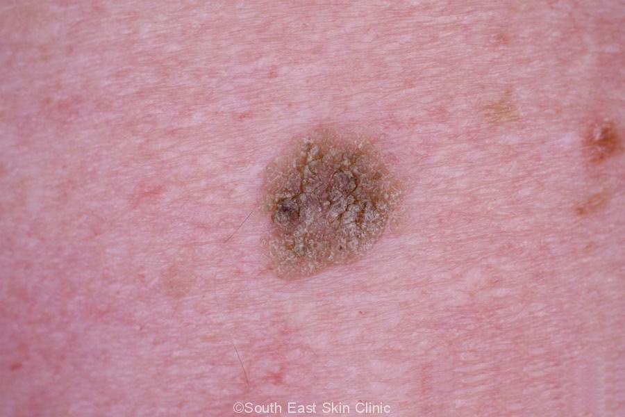
Brown Seborroeic Keratosis
Please click on the images for details.
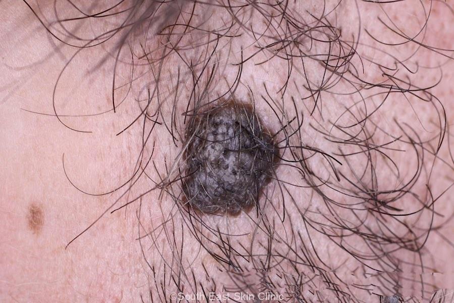
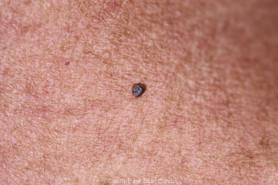
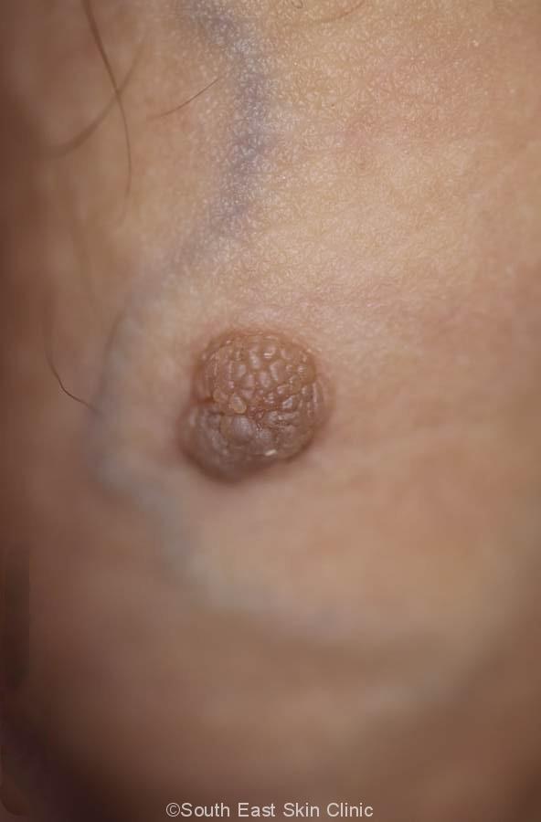
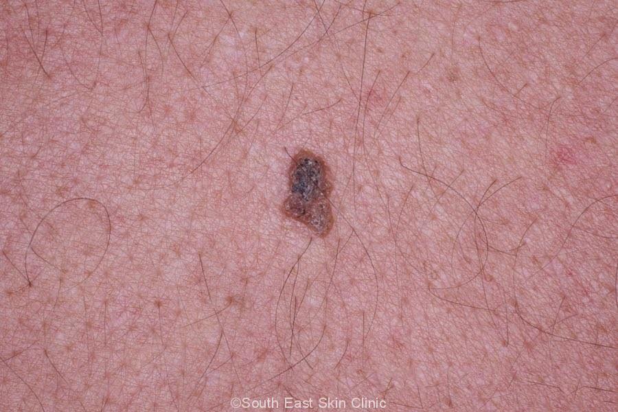
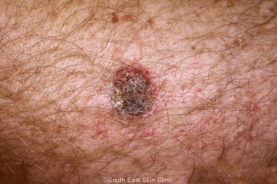
Thicker & Darker Seborroeic Keratosis
Please click on the images for details.
The big issue is really that SK may occasionally mimic a melanoma.
Seborrheic Keratosis does not require treatment unless there are cosmetic concerns or the lesion catches on things like clothing. In this scenario, a medicare rebate may not be available unless a biopsy ‘is clinically necessary to confirm the diagnosis for appropriate management of the lesion.’ There are certain occasions, however, when a biopsy is required to distinguish a seborrhoeic keratosis from a melanoma or another type of skin lesion.
This SK on the side of the face was both unsightly and caught on a shaver:
Treatment options will depend on the size and location of the lesion.
Usual treatment options are:
A common question is ‘What can I do to stop getting them?’
Traditionally, you either put up with them or arrange to have them removed as a cosmetic procedure. There is some evidence that ammonium lactate or tazarotene 0.1% cream may reduce the height or improve the appearance of individual lesions.
Unfortunately, there is currently no treatment that has been truly proven to work. There will surely be a topical product in the future that will work – but when this will be available is anyone’s guess. Research has recently found that the lesions ‘are sensitive to inhibition by ATP-competitive Akt inhibitors’, which should lead to new topical products.
Leser-Trélat described The sudden eruption of large numbers of inflamed SKs in association with internal malignancy. This has been described in case reports, and it is still not known whether this is truly an association. However, it usual to run some cancer tests in younger people with the sudden eruption of multiple SKs.
Everyone over 40 years of age has at least one ‘SK.’
 ©South East Skin Clinic, All rights reserved
 ©South East Skin Clinic, All rights reservedMole (Nevus)
